

MEDIHUB STROKE
AI Stroke Total Solution

MEDIHUB STROKE
Stroke Total Solution

Fast. Accurate. Healthy.
Stroke Total Solution
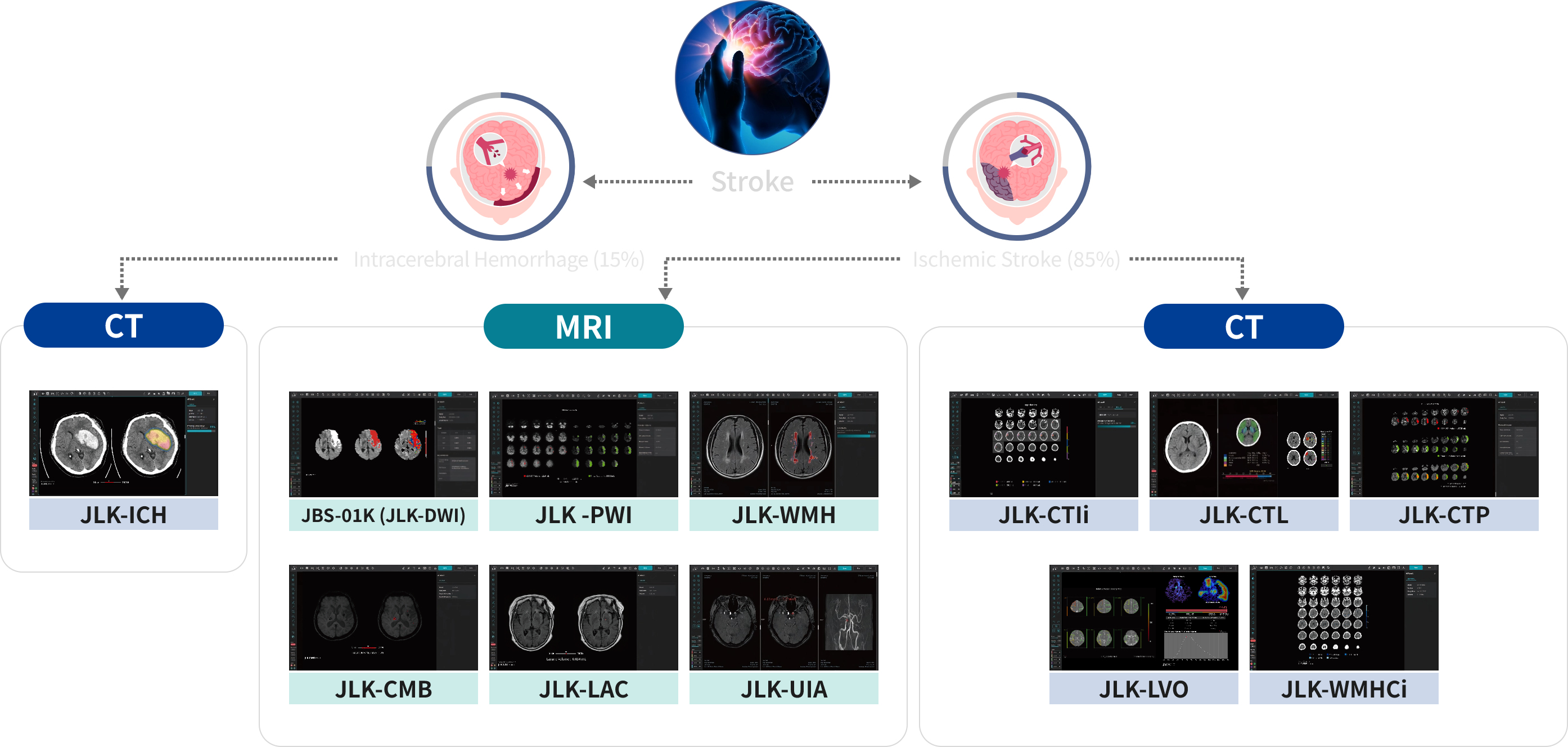
Stroke can be classified into two categories: Hemorrhage and ischemia. Stroke is a serious disorder that occurs commonly so fast and accurate treatment within golden hour is crucial. MEDIHUB STROKE assists fast and accurate stroke diagnosis system in a single platform with various medical image (CT, CTA, MRI, MRA) analysis technology.

Hemorrhagic stroke is a cerebrovascular disorder caused by
ruptures of cerebral blood vessels.
The common causes could be high blood pressure, brain aneurysm
ruptures, etc.
Emergency hemorrhagic stroke patients are immediately diagnosed
via CT images and in this step, JLK's flagship solutions, JLK-ICH
and JLK-CTIi detects and marks the presence of dark areas in regions excluding the cerebrospinal fluid area within brain CT images.
Ischemic stroke occurs more frequently than hemorrhagic strokes
(83%) and it is a disorder with blocked blood vessels that cause
part of the brain to be damaged.
The damaged part where blood vessels were is necrotized, leaving
symptoms permanently.
Patients with suspected cerebral infarction are found more easily
in MRI after a CT scan.
MEDIHUB STROKE's MRI solutions assist faster and more accurate
treatment methods by offering quantitative information on the
diagnosis and treatment of acute ischemic strokes.
AI Solution Pipeline
-
-
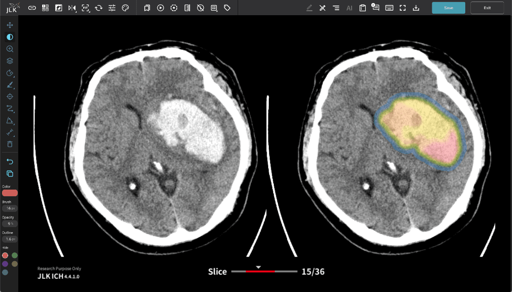
JLK-ICH
NCCT IntracranialHemorrhageDetection SubtypeClassification
AI Based Hemorrhagic Stroke Detection Solution
JLK-ICH is an AI-based medical solution that analyzes brain CT images to detect the presence and location of intracranial hemorrhage.
This solution was developed to rapidly analyze the presence of cerebral hemorrhage in emergency situations and assists medical staff in making quick decisions and diagnoses.
-
JLK-ICH
-
-
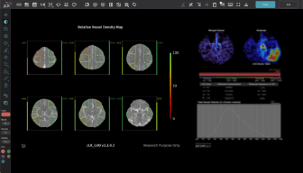
JLK-LVO
CT Angiography LVOdetection DensityComparison
AI Based Large Vessel Occlusion Detection Solution
JLK-LVO uses AI to analyze CT angiography images and provides information on suspected large vessel occlusion areas. Based on the analyzed results, it provides cerebral vascular density comparison information for bilateral middle cerebral artery regions.
The provided information assists medical staff in diagnosis and treatment decisions and can help in patient-specific treatment planning.
-
JLK-LVO
-
-
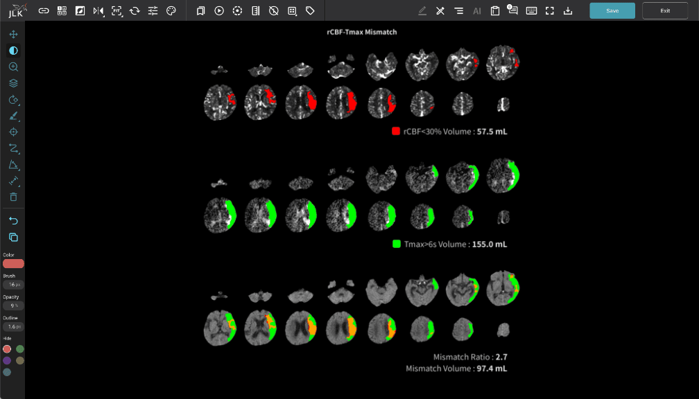
JLK-CTP
CT Perfusion Tmax>6s rCBF<30% MismatchVolume&Ratio
Brain Computed Tomography Perfusion Image Analysis Solution
JLK-CTP analyzes brain CT perfusion images to automatically calculate the volume of the infarct core and the volume of perfusion deficit areas.
Based on this, it measures the volume of the penumbra to assist in determining treatment plans for hyperacute ischemic stroke.
-
JLK-CTP
-
-
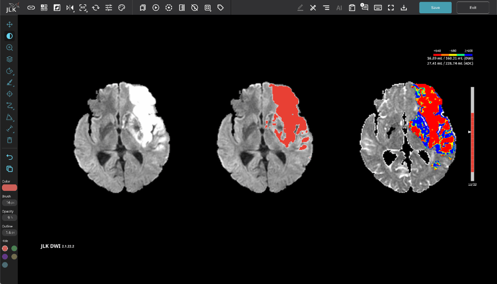
JLK-DWI
MR Diffusion IschemicStrokeSubtype LesionDetection
AI-Powered Ischemic Stroke Subtype Analysis Software
JLK-DWI uses AI to detect suspected cerebral infarction lesions in diffusion-weighted MR images and analyzes the size, location, and pattern of the lesions.
Based on the analyzed results, it provides probability values for large-artery atherosclerosis, cardioembolism, and small-vessel occlusion to assist medical staff in diagnosis and treatment.
-
JLK-DWI
-
-
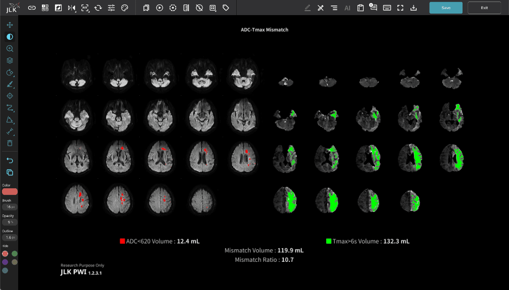
JLK-PWI
MR Perfusion Tmax>6s rCBF<30% MismatchVolume&Ratio
Brain Magnetic Resonance Perfusion Image Analysis Solution
JLK-PWI automatically calculates the infarct core volume based on diffusion-weighted MR images and automatically calculates the volume of perfusion deficit areas based on perfusion images.
Based on this, it measures the volume of the penumbra to assist in determining treatment plans for hyperacute ischemic stroke.
-
JLK-PWI
-
-
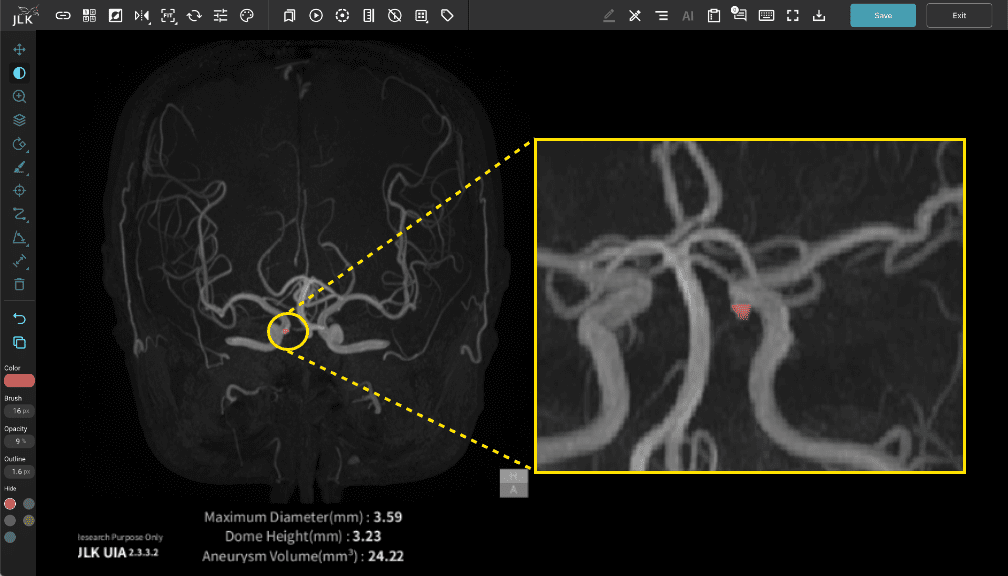
JLK-UIA
3D TOF MRA AneurysmDetection AneurysmAnalysis
AI Based Cerebral Aneurysm Detection Solution
JLK-UIA analyzes time-of-flight images from brain magnetic resonance angiography to detect suspected unruptured intracranial aneurysm areas and provides morphological information such as the size and volume of aneurysms.
The provided information assists medical professionals in diagnosis.
-
JLK-UIA